The Centre for Bone and Periodontal Research Français
Centre for Bone and Periodontal Research
Histology
Imaging and Biomechanics
Biochemistry
Imaging and Biomechanics

Micro Computed Tomography

Representative Applications

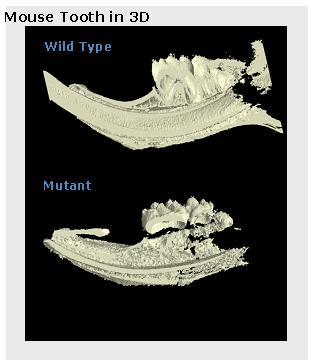
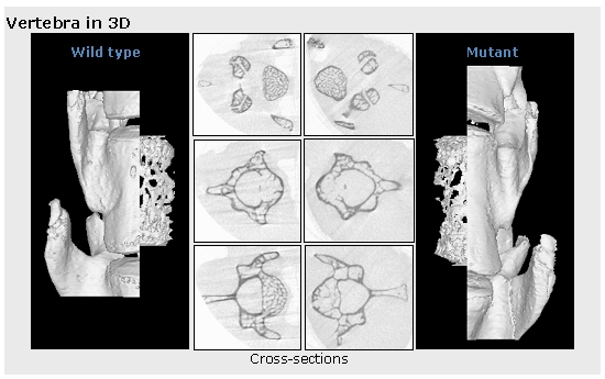
Quantitation of trabecular bone 3d architecture:

  • Bone Volume
  • Bone surface
  • SMI (structure model index)
  • Trabecular pattern factor, thickness and separation
  • Porosity
  • BMD

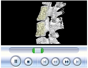
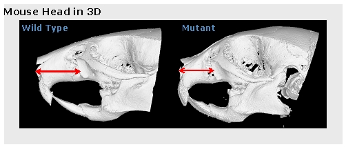
Ex.: Limb, vertebrae, cranial vault and jaw.


3d and 2d Visualization:
  • 3D images and slides
  • 2D cross sections

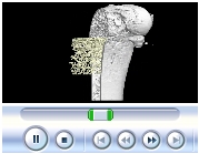
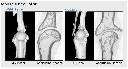
Ex.: Shoulder and knee joint or tibia and mandible regeneration.


Animations:
  • Showing inner
  • micro structure
  • Showing outer surface

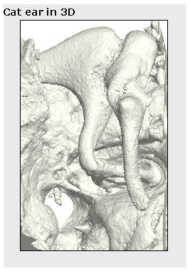
Ex.: Cat inner ear, dog palate regeneration or bone cement.


Instrumentation

Micro-CT technology enables investigators to nondestructively obtain in-depth 3D structure and quantitative data of hard and soft tissues of small animals and other composite materials as well.

Specifications

  • Energy range: 20 to 100kV
  • Current range: 0 to 250µA
  • Resolution range: 0.8 to 25µm
  • Detector: 8000 x 8000 pixels 12-bit cooled CCD fiber-optics camera coupled to scintillator
  • Maximum sample size : < 50 mm

Results

3D Femur Animation
3D Femur Animation
3D Spine Animation
3D Spine Animation

Mouse Tooth in 3D
Mouse Tooth in 3D
Cat Ear in 3D
Cat Ear in 3D
Mouse Knee Joint
Mouse Knee Joint
Mouse Head in 3D
Mouse Head in 3D
Vertebra in 3D
Vertebra in 3D
Neo Natal Mouse Skull


Imaging/Biomechanics Technologies

Micro Computed Tomography
Bone Densitometry
Radiography
Small Animal Live Imaging
Biomechanical Testing


Download PDF Forms

New Investigator
New Project
Imaging/Biomechanics Service Request


Contact

Yongjun Xiao, Ph.D.
Tel:  514 398-4480
Fax:  514 398-4020
Email:  Yongjun.xiao@
mail.mcgill.ca


McGill University   Université de Montréal   École Polytechnique   Centre Hospitalier de l'Université de Montréal   Institut de Recherches Cliniques de Montréal