|
|
Imaging and Biomechanics
Small Animal Live Imaging
Representative applications

The eXplore Optix is a live animal (rat and mouse) optical imaging system that permits the detection
of light emitted in the visible and near-infrared region of the spectrum. Applications include monitoring
tumor growth and metastasis, the distribution of cell populations in the animal (eg, immune cells), in
vivo gene expression of engineered reporters, injection of antibodies, growth factors or peptides conjugated to
fluorophores that can be used to localize receptor expression or monitor drug distribution, absorption and clearance.
Instrument
This system has the potential for multi-wavelength imaging with the addition of multiple lasers
(the ability to image in the Near-Infrared region (650-900nm) will enhance the sensitivity of the system
due to better tissue penetration of longer wavelengths). The system collects fluorescent intensity data
and calculates fluorescence life-time values that permits the identification of specific signals produced
by the fluorescent probe of interest, imaging multiple fluorophores in the same animal and distinguishes
specific signals versus background auto-fluorescence.
Specifications
|
Pulsed laser wavelength: 445nm
Detection wavelength: 445nm (absorption) and 500-550nm (emission)
Detection spot: 1mm diameter
Spatial resolution: 0.5-3mm steps
Scan area: 20x8.4cm
Scan time: adjustable
Animal plate temperature: 26-420C
|

eXplore Optix™ manufactured by ART Inc. and distributed by GE Healthcare Bio-Sciences
|
Results
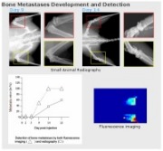
Bone Metastases Development and Detection
Courtesy of Dr. Richard Kremer, MUHC, McGill University
|

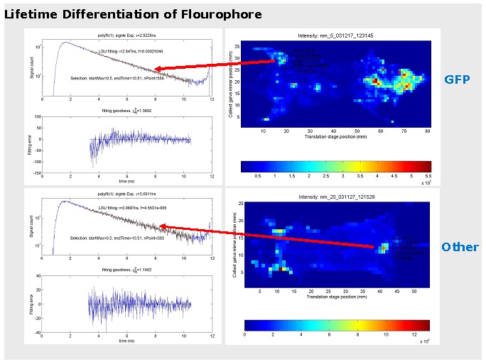
Lifetime Differentiation of Flourophore
Courtesy of Dr. Richard Kremer, MUHC, McGill University
|
|
Imaging/Biomechanics Technologies

Imaging/Biomechanics Galleries
 Photo Gallery Photo Gallery
Equipment Gallery
Download PDF Forms 
|