|
|
Imaging and Biomechanics
Bone Densitometry
Representative Applications

- Mouse live imaging
- Femur/tibia/vertebra ROI
- Ex vivo imaging: canine palate
- Ex vivo imaging: rabbit knee/mandible
Instrumentation
This dedicated small animal densitometer enables monitoring over time of live mice, as
well as ex vivo studies requiring assessment of bone mineral density. Multiple Regions of
Interest (ROI) can be described on each sample. Results are provided in terms of bone
mineral density (g/cm2), bone mineral content (g), fat and lean tissue content (g).
Specifications
- Imaging area: 100 x 80mm
- Focal spot size: 0.25 x 0.25mm
- Energy: 80kV
- Current: 400µA
|
 |
Results
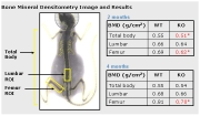
Bone Mineral Densitometry Image and Result
|
|
|
Imaging/Biomechanics Technologies

Imaging/Biomechanics Galleries
 Photo Gallery Photo Gallery
Equipment Gallery
Download PDF Forms 
|